 +1 302-235-9792
 SIGN IN
 CONTACT US


- News
- Fc-Fusion Protein vs PEGylation: Comparative Strategies for Extending the In Vivo Half-Life of Therapeutic Enzymes
share


Fc-Fusion Protein vs PEGylation: Comparative Strategies for Extending the In Vivo Half-Life of Therapeutic Enzymes
Content Menu
● Fc-Fusion Protein vs PEGylation at a Glance
● How Fc Fusion Extends Enzyme Half-Life
>> Why Fc Architecture Matters
>> Linker Design Is a Functional Variable
>> Fc Effector Functions
● How PEGylation Extends Enzyme Half-Life
>> Random PEGylation
>> Site-Specific PEGylation
● Catalytic Activity Versus Circulating Exposure
● Tissue Distribution and Molecular Size
● Developability Risks
>> Activity Loss
>> Aggregation and Molecular Stability
>> Immune-Response Considerations
● Analytical Strategy for Comparing Both Formats
● A Head-to-Head Experimental Workflow
● Practical Selection Matrix
>> Consider Fc Fusion When
>> Consider PEGylation When
>> Consider Parallel Evaluation When
● Expert Design Insights
● Research Support from Gene Universal
● Frequently Asked Questions
>> 1. Is Fc Fusion Always Better Than PEGylation?
>> 2. Does PEGylation Always Reduce Enzyme Activity?
>> 3. Why Do Fc-Fusion Formats Have Different Half-Lives?
>> 4. What PEG Size Should Be Evaluated First?
>> 5. Which Assays Are Essential for an Early Comparison?
>> 6. Can Fc Fusion and PEGylation Be Combined?
>> 7. Should Researchers Select a Strategy Based on Half-Life Data Alone?
● References
Fc-fusion protein vs PEGylation is a critical comparison for researchers seeking to extend the in vivo half-life of therapeutic enzymes. Both strategies can reduce rapid clearance, increase systemic exposure, and protect vulnerable proteins from degradation. However, they achieve these goals through fundamentally different mechanisms.
Fc fusion is a genetically encoded approach. It combines an enzyme with the Fc region of an immunoglobulin, enabling increased molecular size and interaction with the neonatal Fc receptor, or FcRn. PEGylation is a post-expression modification in which polyethylene glycol chains are covalently attached to the enzyme to increase its hydrodynamic volume and create a protective steric barrier.
The best choice cannot be determined by half-life alone. Researchers must also consider catalytic activity, molecular architecture, expression feasibility, conjugation efficiency, tissue access, aggregation, immune-response risk, and analytical complexity.
For many enzyme programs, the most reliable strategy is to create a small, balanced panel of Fc-fusion and PEGylated variants and compare them using fit-for-purpose research-grade materials.
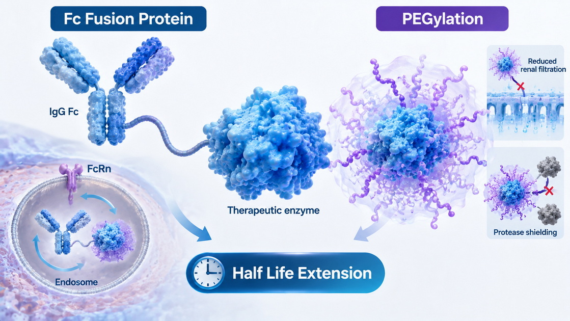
Fc-Fusion Protein vs PEGylation at a Glance
| Decision factor | Fc-fusion protein | PEGylation |
|---|---|---|
| Primary mechanism | FcRn-mediated recycling and increased molecular size | Increased hydrodynamic size, steric shielding, and reduced renal filtration |
| Production method | Genetically encoded fusion construct | Chemical or enzymatic modification after protein production |
| Typical architecture | Often dimeric, depending on Fc format | Linear or branched polymer attached at one or more sites |
| Molecular consistency | Potentially high when sequence, assembly, and glycosylation are controlled | Depends heavily on conjugation method and attachment-site control |
| Main activity risk | Fusion orientation or linker may interfere with enzyme structure | PEG may block the active site or restrict substrate access |
| Expression implications | Requires correct folding, assembly, and Fc glycosylation | Requires expression of the parent enzyme followed by conjugation |
| Important attributes | FcRn binding, Fc receptor interactions, glycans, aggregation, linker integrity | PEG occupancy, attachment sites, free PEG, unconjugated enzyme, positional isomers |
| Common advantage | Genetically defined architecture with receptor-mediated recycling | Strong steric shielding without adding another protein domain |
| Important uncertainty | Cargo and Fc architecture can alter FcRn recycling | Anti-PEG antibodies and heterogeneous conjugation can affect exposure |
The comparison shows that Fc fusion is not merely a molecular-weight strategy, while PEGylation is not simply a method for coating a protein. Each format changes how the enzyme behaves as a complete molecular system.
How Fc Fusion Extends Enzyme Half-Life
An Fc-fusion protein contains a functional protein domain joined to the Fc region of an immunoglobulin. In enzyme applications, the catalytic protein may be attached to either the N-terminus or C-terminus of the Fc through a designed peptide linker.
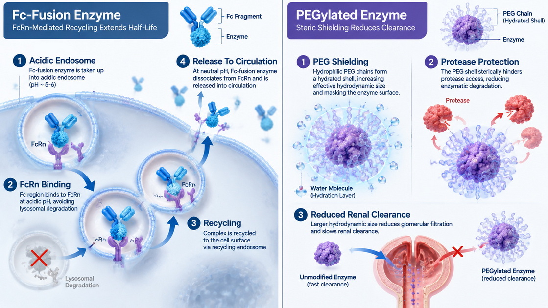
The Fc region can interact with FcRn inside acidic endosomes. After a circulating protein is internalized through nonspecific cellular uptake, FcRn can bind the Fc portion and redirect the molecule away from lysosomal degradation.
The Fc-fusion protein is then transported back to the cell surface. At the near-neutral pH outside the cell, the interaction weakens, allowing the protein to return to circulation.
This mechanism can provide two half-life-extension effects:
- Increased molecular size, which may reduce rapid renal filtration.
- FcRn-mediated recycling, which can protect the molecule from intracellular degradation.
However, adding an Fc region does not guarantee that an enzyme will acquire the same circulation time as a full-length IgG. The fusion partner can influence molecular charge, FcRn access, endosomal trafficking, aggregation, target-mediated clearance, and release from FcRn at neutral pH.
Why Fc Architecture Matters
Many Fc-fusion constructs naturally form homodimers through interactions in the Fc region. Dimerization can be beneficial if the enzyme functions as a multimer or benefits from increased avidity. It can be harmful if the enzyme requires a particular oligomeric arrangement.
A dimeric Fc fusion may also produce unwanted spatial constraints. Two enzyme domains positioned close together can interfere with substrate access or promote non-native interactions.
Researchers should therefore evaluate factors such as:
- Monovalent versus divalent enzyme presentation.
- Homodimeric versus heterodimeric Fc architectures.
- N-terminal versus C-terminal fusion.
- Linker length and flexibility.
- Fc isotype and Fc engineering.
- Distance between the enzyme and Fc domain.
A recent study of the IgG-degrading enzyme IdeS illustrates this point. Both monovalent and divalent Fc-fusion formats retained IgG-cleaving activity, although their kinetics differed from those of the native enzyme. The monovalent format achieved an approximately sevenfold half-life extension in mice, demonstrating that architecture can determine whether Fc attachment translates into longer functional exposure.
Linker Design Is a Functional Variable
The linker between the enzyme and Fc should not be treated as an inactive spacer. It can influence folding, expression, catalytic accessibility, stability, and proteolytic sensitivity.
A very short linker may create steric interference between the Fc and enzyme domains. A long flexible linker can improve domain mobility but may increase susceptibility to cleavage or permit unfavorable intramolecular interactions.
Common experimental variables include:
- Flexible glycine-serine-rich linkers.
- Semi-rigid helical linkers.
- Protease-resistant sequences.
- Linkers with different lengths.
- Linkers designed to reduce domain-to-domain interference.
The preferred linker is not necessarily the longest or most flexible one. It is the linker that delivers the best balance of enzyme activity, expression yield, molecular stability, assembly, and FcRn-dependent behavior.
Fc Effector Functions
The Fc domain may interact with Fc gamma receptors or complement-associated proteins. These interactions are not always desirable when Fc is used primarily as a half-life-extension module.
Fc engineering can reduce selected effector interactions. However, every modification should be evaluated experimentally because changes in Fc sequence can influence thermal stability, glycosylation, receptor binding, and pharmacokinetic behavior.
How PEGylation Extends Enzyme Half-Life
PEGylation is the covalent attachment of one or more polyethylene glycol chains to a protein. PEG is highly hydrophilic and attracts water molecules, creating a hydrated molecular envelope around the modified enzyme.
This envelope makes the conjugate behave as if it were larger than its protein mass alone would suggest. The increased hydrodynamic volume can reduce glomerular filtration and slow renal clearance.
PEG may also provide several additional effects:
- Reduced access by proteolytic enzymes.
- Lower nonspecific protein interactions.
- Increased apparent solubility.
- Reduced aggregation in some formulations.
- Partial shielding of exposed protein epitopes.
These benefits are not automatic. The same PEG layer that protects an enzyme can also limit substrate diffusion, interfere with receptor binding, or reduce cellular uptake.
The optimal design is therefore not the conjugate with the greatest number of PEG chains. It is the design that provides adequate protection while retaining sufficient catalytic function.
Random PEGylation
Traditional PEGylation often uses amine-reactive chemistry. This approach can modify the N-terminus and accessible lysine residues on the protein surface.
If the enzyme contains many exposed lysines, random modification may generate a complex mixture that includes:
- Unmodified protein.
- Mono-PEGylated protein.
- Multi-PEGylated protein.
- Positional isomers.
- Molecules with different catalytic activities.
- Molecules with different stability or clearance behavior.
The average PEG-to-protein ratio does not fully describe such a mixture. Two conjugates with the same overall PEG occupancy may contain different attachment patterns and biological properties.
Site-Specific PEGylation
Site-specific PEGylation places the polymer at a deliberately selected position. Ideally, this position is exposed, chemically accessible, and separated from the active site, substrate channel, receptor-binding region, and oligomerization interface.
Possible methods include:
- Selective N-terminal PEGylation.
- Engineered cysteine conjugation.
- Enzyme-mediated conjugation.
- Noncanonical amino-acid incorporation.
- Chemoselective modification of an introduced functional group.
Site-specific attachment can produce a more defined conjugate and improve structure–function interpretation. It can also reduce the need to separate numerous positional isomers.
Published L-asparaginase research provides a useful example. A site-specific monoPEGylated format retained an initial specific activity of approximately 233 U/mg. A randomly polyPEGylated preparation in the same study retained approximately 21% of the specific activity of the unmodified enzyme.
This does not mean that random PEGylation always produces unacceptable activity. It shows that attachment position and modification level may be more important than PEGylation itself.
Catalytic Activity Versus Circulating Exposure
Half-life should never be evaluated as an isolated number. Therapeutic enzymes perform catalytic reactions, meaning that their value depends on retained function as well as molecular concentration.
A modified enzyme may remain detectable in plasma while gradually losing catalytic activity. Concentration-based assays could interpret this as persistent exposure, even though the circulating molecules are no longer functionally active.
A stronger experimental plan measures both:
- Protein concentration over time.
- Residual catalytic activity over time.
The relationship between these measurements reveals whether longer molecular persistence corresponds to longer functional persistence.
Early studies should also examine:
- Michaelis constant and maximum reaction velocity.
- Specific activity per milligram or per mole.
- Substrate selectivity.
- Inhibitor sensitivity.
- Activity in serum or plasma.
- Activity after freeze–thaw cycles.
- Activity following prolonged incubation.
- Protease resistance.
For enzymes with very small substrates, a PEG layer may still allow sufficient substrate diffusion. For enzymes acting on large proteins or sterically restricted substrates, PEG shielding may have a greater functional effect.
Tissue Distribution and Molecular Size
Increasing molecular size can reduce renal filtration, but it may also limit tissue access. This trade-off applies to both Fc fusion and PEGylation.
Fc fusion adds a substantial protein domain and frequently produces a dimeric molecule. PEG adds less true molecular mass in some designs but creates a comparatively large hydrated volume.
The consequences depend on where the enzyme must act. A format optimized for persistence in blood may not be ideal when the enzyme must reach:
- Dense extracellular matrix.
- Poorly vascularized tissue.
- Large deposited substrates.
- Intracellular compartments.
- Restricted anatomical spaces.
- Cell-surface targets with limited accessibility.
Researchers should therefore include tissue-distribution requirements in the initial design plan rather than evaluating distribution after selecting a format.
Developability Risks
Activity Loss
Fc fusion may reduce activity by altering enzyme orientation, oligomerization, mobility, or access to the catalytic site. PEGylation may reduce activity through steric shielding, modification of functional residues, or restriction of conformational changes required for catalysis.
Structural mapping should be completed before selecting fusion junctions or conjugation sites.
Aggregation and Molecular Stability
Fc-fusion proteins may aggregate because of domain misfolding, exposed hydrophobic surfaces, incorrect assembly, or unfavorable enzyme–Fc interactions. Their glycosylation and charge profiles may create additional heterogeneity.
PEGylation can improve apparent solubility, but the conjugation reaction may introduce partially modified species, residual reagents, aggregates, or unstable conjugates.
Both strategies require orthogonal analysis rather than dependence on a single purity method.
Immune-Response Considerations
Immune responses to protein-based materials can be influenced by sequence, structure, aggregates, process impurities, formulation, administration route, and exposure pattern.
Fc fusion can introduce new structural features at the fusion junction. The linker, Fc region, altered oligomeric state, or partially unfolded domains may also influence recognition.
PEG can shield protein epitopes, but antibodies against PEG may be present before exposure or develop following administration. Such antibodies may influence clearance, exposure, or assay interpretation.
A PEG-related assessment may need to distinguish antibodies directed against:
- The enzyme component.
- The PEG component.
- The complete PEG–enzyme conjugate.
- Newly formed structural features near the attachment site.
For an Fc fusion, the corresponding assessment may need to distinguish responses to the enzyme, linker, Fc domain, and fusion junction.
Analytical Strategy for Comparing Both Formats
A balanced comparison should use equivalent analytical depth for each format.
| Attribute | Fc-fusion assays | PEGylated-enzyme assays |
|---|---|---|
| Identity | Peptide mapping and intact mass | Peptide mapping, intact mass, and conjugation-site mapping |
| Size variants | SEC, light scattering, electrophoresis | SEC, light scattering, and separation by PEG occupancy |
| Functional activity | Catalytic kinetics and substrate testing | Catalytic kinetics and substrate testing |
| Mechanism-specific testing | FcRn binding and pH-dependent release | PEG occupancy and attachment-site confirmation |
| Molecular heterogeneity | Glycan profile, charge variants, assembly | Positional isomers, free PEG, and unconjugated enzyme |
| Stability | Thermal, chemical, and serum stability | Thermal, chemical, conjugate, and serum stability |
| Immune-risk research | Enzyme-, linker-, and Fc-related assessments | Enzyme-, PEG-, and conjugate-related assessments |
A single analytical method rarely provides sufficient information. Size-exclusion chromatography may detect aggregates but cannot fully characterize conjugation sites, Fc glycosylation, charge variants, or catalytic integrity.
A Head-to-Head Experimental Workflow
A practical comparison can be organized into eight steps.
1. Define the desired profile. Establish the required activity, exposure duration, route, tissue compartment, concentration range, and acceptable molecular complexity.
2. Map structural constraints. Identify catalytic residues, substrate channels, oligomerization interfaces, disulfide bonds, glycosylation sites, and accessible conjugation positions.
3. Create a balanced design panel. Compare multiple Fc orientations or linkers with several PEG sizes or attachment sites.
4. Use a consistent enzyme background. Begin with the same enzyme sequence and comparable purification standards to avoid confounding variables.
5. Evaluate expression and recovery. Measure soluble expression, purification yield, assembly, purity, and batch-to-batch consistency.
6. Measure catalytic function. Determine kinetic parameters, specific activity, serum stability, and activity following stress conditions.
7. Assess mechanism-specific attributes. Examine FcRn binding and release for Fc constructs. Determine PEG occupancy, attachment sites, free PEG, and residual parent enzyme for PEGylated constructs.
8. Compare exposure with function. Evaluate both circulating concentration and residual catalytic activity over time in an appropriate research model.
Practical Selection Matrix
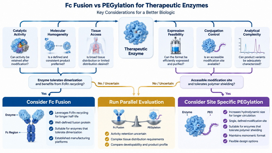
Consider Fc Fusion When
- A genetically encoded and sequence-defined construct is preferred.
- The enzyme tolerates increased size or dimerization.
- FcRn-mediated recycling is a central design objective.
- Mammalian expression can support appropriate folding and assembly.
- Fc interactions can be measured and adjusted when necessary.
- Site-specific chemical conjugation is difficult to implement.
Consider PEGylation When
- The parent enzyme expresses and purifies efficiently.
- A suitable surface attachment site can be identified.
- Steric protection and reduced renal filtration are primary goals.
- The enzyme tolerates the selected PEG architecture.
- A defined conjugation process can produce a sufficiently consistent material.
- The research plan can characterize PEG-related species.
Consider Parallel Evaluation When
- The enzyme has a narrow substrate channel.
- Oligomerization strongly influences activity.
- Tissue access is as important as plasma persistence.
- Fc-induced dimerization may alter function.
- No validated PEG attachment site is available.
- The preferred strategy depends mainly on assumptions rather than comparative data.
Expert Design Insights
A common mistake is to optimize the longest measurable half-life rather than the longest functional half-life. For catalytic proteins, substrate turnover and retained activity may be more informative than total circulating concentration.
Another mistake is to compare one highly optimized format with one preliminary format. A carefully selected site-specific PEG conjugate should not be compared with the first available Fc construct. Likewise, an optimized Fc architecture should not be compared with an uncontrolled PEGylation mixture.
The comparison should be design-balanced. Similar effort should be applied to variant selection, purification, analytical characterization, and functional testing.
Finally, the analytical plan should follow the extension mechanism. Fc fusion requires focused attention to FcRn behavior, assembly, glycans, linker integrity, and Fc-mediated interactions. PEGylation requires detailed characterization of attachment sites, PEG occupancy, free polymer, residual parent enzyme, and positional heterogeneity.
Research Support from Gene Universal
Gene Universal provides global research support across DNA/RNA services, recombinant protein expression, antibody expression, purification, and related early discovery and characterization workflows.
For teams comparing Fc-fusion proteins and PEGylated enzymes, a productive starting point may include:
- Fc-fusion construct and linker design.
- Sequence optimization.
- Expression-host evaluation.
- Small-scale expression screening.
- Recombinant enzyme production.
- Parent-enzyme purification.
- Research-use analytical characterization.
- Production of materials for comparative laboratory studies.
Gene Universal provides research-use candidates, fit-for-purpose research-grade materials, and preclinical research support. Its services do not include GMP manufacturing, CDMO programs, or IND submission support.
Planning an Fc-fusion protein panel or preparing a parent enzyme for PEGylation studies? Contact Gene Universal to discuss construct design, expression screening, purification, and research-use characterization tailored to your enzyme program.
Frequently Asked Questions
1. Is Fc Fusion Always Better Than PEGylation?
No. Fc fusion provides FcRn-mediated recycling, but the additional size, dimerization, glycosylation, and Fc-associated interactions may alter enzyme behavior. PEGylation provides strong steric shielding, but conjugation can reduce activity or generate heterogeneous species.
2. Does PEGylation Always Reduce Enzyme Activity?
No. Activity retention depends on the PEG size, architecture, attachment site, degree of modification, and distance from functional surfaces. Site-specific conjugation away from the active site can preserve a high proportion of catalytic activity.
3. Why Do Fc-Fusion Formats Have Different Half-Lives?
Fusion orientation, linker properties, valency, charge, aggregation, FcRn-binding kinetics, target-mediated clearance, and the enzyme cargo itself can alter disposition. The Fc domain provides a recycling mechanism, but the complete molecular architecture determines the result.
4. What PEG Size Should Be Evaluated First?
There is no universally optimal size. Researchers can compare several linear or branched PEG formats while keeping the attachment site consistent. The preferred format should provide adequate shielding without excessive loss of catalytic activity or tissue access.
5. Which Assays Are Essential for an Early Comparison?
Start with identity, purity, aggregation, intact mass, thermal stability, serum stability, and catalytic kinetics. Add FcRn-binding analysis for Fc fusions and conjugation-site mapping, PEG occupancy, free PEG, and residual parent-enzyme measurements for PEGylated formats.
6. Can Fc Fusion and PEGylation Be Combined?
Yes, the two approaches can be combined experimentally. However, dual modification increases molecular and analytical complexity. It should only be considered when each modification serves a defined purpose that cannot be achieved with a simpler design.
7. Should Researchers Select a Strategy Based on Half-Life Data Alone?
No. Selection should consider functional activity, tissue distribution, molecular stability, expression or conjugation yield, aggregation, analytical tractability, and immune-response hypotheses. The longest plasma half-life may not produce the longest period of useful catalytic activity.
References
1. Strohl WR. [Fusion Proteins for Half-Life Extension of Biologics as a Strategy to Make Biobetters]. *BioDrugs*. 2015;29(4):215–239.
2. Ebrahimi SB, Samanta D. [Engineering Protein-Based Therapeutics through Structural and Chemical Design]. *Nature Communications*. 2023;14:2411.
3. Unverdorben F, Richter F, Hutt M, et al. [Pharmacokinetic Properties of IgG and Various Fc Fusion Proteins in Mice]. *mAbs*. 2016;8(1):120–128.
4. Chen X, Zaro JL, Shen WC. [Fusion Protein Linkers: Property, Design and Functionality]. *Advanced Drug Delivery Reviews*. 2013;65(10):1357–1369.
5. Meneguetti GP, Santos JHPM, Obreque KMT, et al. [Novel Site-Specific PEGylated L-Asparaginase]. *PLOS ONE*. 2019;14(2):e0211951.
6. Kozma GT, Shimizu T, Ishida T, Szebeni J. [Anti-PEG Antibodies: Properties, Formation, Testing and Role in Adverse Immune Reactions to PEGylated Nano-Biopharmaceuticals]. *Advanced Drug Delivery Reviews*. 2020;154–155:163–175.
7. Wei Y, et al. [Half-Life Extension of the IgG-Degrading Enzyme IdeS Using Fc Fusion]. 2025.
8. Wu B, Sun YN. [Pharmacokinetics of Peptide-Fc Fusion Proteins]. *Journal of Pharmaceutical Sciences*. 2014;103(1):53–64.
9. U.S. Food and Drug Administration. [Immunogenicity Assessment for Therapeutic Protein Products]. Guidance for Industry.
10. U.S. Food and Drug Administration. [Immunogenicity Testing of Therapeutic Protein Products—Developing and Validating Assays for Anti-Drug Antibody Detection]. Guidance for Industry.
11. European Medicines Agency. [Immunogenicity Assessment of Biotechnology-Derived Therapeutic Proteins]. Scientific Guideline.