 +1 302-235-9792
 SIGN IN
 CONTACT US


- News
- Periplasmic Secretion Vs Whole-Cell Lysis: Minimizing Proteolytic Degradation of Small Bioactive Peptides in E Coli
share


Periplasmic Secretion Vs Whole-Cell Lysis: Minimizing Proteolytic Degradation of Small Bioactive Peptides in E Coli
Content Menu
● Why Small Bioactive Peptides Are Vulnerable in E. coli
>> The Main Sources of Peptide Loss
● Periplasmic Secretion for Small Bioactive Peptides
>> Why the Periplasm Can Be Advantageous
>> Key Limitations of Periplasmic Secretion
>> When Periplasmic Secretion Is Often the Better Starting Point
● Whole-Cell Lysis for Intracellular Peptide Recovery
>> Why Whole-Cell Lysis Remains Valuable
>> The Main Proteolysis Challenge After Lysis
>> How to Make Whole-Cell Lysis More Protective
● Periplasmic Secretion vs Whole-Cell Lysis: Direct Comparison
● A Practical Decision Workflow for Peptide Expression
>> 1: Assess the Peptide Sequence
>> 2: Build a Small, Purposeful Construct Panel
>> 3: Measure Intact Product, Not Just Apparent Yield
>> 4: Use Time-Course Sampling
>> 5: Select the Route Based on Total Usable Recovery
● Reducing Proteolysis Before It Becomes a Downstream Problem
>> Design for Protection, Then Release
>> Avoid Overdriving Expression
>> Treat Signal Peptides as Screening Variables
>> Confirm Sequence Integrity Early
● Conclusion
● Frequently Asked Questions
>> 1. Is periplasmic secretion always better than whole-cell lysis for small peptides?
>> 2. Why are small bioactive peptides more susceptible to proteolytic degradation?
>> 3. Can a fusion partner reduce peptide degradation in E. coli?
>> 4. Does the E. coli periplasm help form disulfide bonds?
>> 5. How can I determine whether degradation happens during expression or after lysis?
>> 6. Which analytical methods are most useful for checking peptide integrity?
>> 7. Can lower expression temperature reduce peptide degradation?
● References
Small bioactive peptides can be among the most difficult recombinant products to recover from Escherichia coli. Their short length, exposed termini, structural sensitivity, and—in some cases—host-cell toxicity make them particularly vulnerable to proteolytic degradation. For many research programs, the central process-design question is whether to direct the product to the bacterial periplasm or retain it inside the cell for recovery by whole-cell lysis.
Both approaches can support productive peptide expression. However, they create very different biological environments, purification burdens, folding conditions, and degradation risks. In practical protein and antibody expression work, the best route is rarely chosen by yield alone. A useful decision requires evaluating peptide sequence, disulfide-bond requirements, intracellular toxicity, fusion-tag strategy, downstream workflow, and the quality attributes needed for the intended research application.
This article compares periplasmic secretion vs whole-cell lysis for small bioactive peptides in *E. coli*, with a specific focus on reducing peptide loss caused by host proteases and sample-handling stress.
Why Small Bioactive Peptides Are Vulnerable in E. coli
A small bioactive peptide does not behave like a typical soluble enzyme. A larger folded protein may bury potentially susceptible cleavage sites inside a stable three-dimensional structure. In contrast, short peptides often have accessible termini and flexible regions that are easier for proteases to recognize and cleave.
This vulnerability can appear at several stages:
- During intracellular expression, when the peptide is exposed to cytoplasmic proteases.
- During translocation, when export through the inner membrane is incomplete or slow.
- After cell disruption, when cytoplasmic, membrane-associated, and periplasmic components mix together.
- During purification, when long handling times, suboptimal temperature control, or repeated freeze-thaw cycles increase degradation.
- After cleavage from a fusion partner, when the liberated peptide becomes more exposed and less stable.
Historical studies of recombinant secretion in E. coli have noted that smaller polypeptides, including somatostatin, insulin-like growth factor 1, and human epidermal growth factor, may be especially sensitive to intracellular degradation. Directing suitable products toward the periplasm can reduce exposure to certain cytoplasmic proteases while also providing a compartment more favorable for oxidative folding.
For peptide projects, the central challenge is not simply "Can the host express it?" The more useful question is:
The Main Sources of Peptide Loss
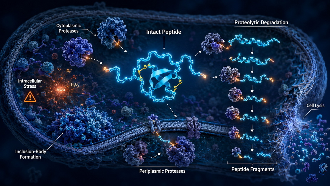
Small bioactive peptides may be lost through more than one mechanism. Identifying the dominant mechanism early helps avoid unnecessary optimization cycles.
| Source of loss | Typical consequence | Why it matters for small peptides |
|---|---|---|
| Cytoplasmic proteases | Truncation or complete degradation | Short, flexible peptides are often accessible substrates |
| Periplasmic proteases | Product clipping after export | Periplasmic targeting reduces some risks but does not eliminate proteolysis |
| Outer-membrane proteases | Cleavage during expression or extraction | Membrane-associated protease activity can affect released material |
| Host toxicity | Poor growth or selection for low-expression variants | Antimicrobial and membrane-active peptides can harm the host |
| Incorrect disulfide pairing | Lower activity, aggregation, heterogeneous forms | Important for cysteine-rich peptides |
| Harsh lysis | Denaturation, aggregation, increased protease exposure | Lysis combines cellular compartments and releases many proteases |
| Slow processing | Progressive degradation after harvest | Small products can degrade before purification is complete |
One important point is often overlooked: the periplasm is not protease-free. *E. coli* has proteolytic activity in multiple cellular locations, including the periplasm, membrane, and cytoplasm. Therefore, periplasmic secretion should be viewed as a risk-management strategy rather than a universal protection mechanism.
Periplasmic Secretion for Small Bioactive Peptides
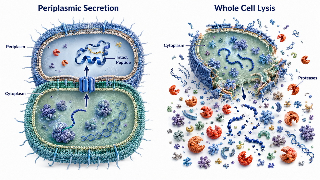
Periplasmic secretion directs the recombinant product across the inner membrane into the space between the inner and outer membranes of Gram-negative bacteria. In most designs, this is achieved by placing an N-terminal signal peptide upstream of the recombinant sequence.
After translocation, the signal peptide is usually removed by host processing machinery. The desired peptide, fusion protein, or peptide precursor can then accumulate in the periplasm or, in some designs, be released further into the culture medium.
Why the Periplasm Can Be Advantageous
The periplasm offers several advantages for research-grade peptide production.
Reduced exposure to cytoplasmic proteases. Cytoplasmic expression places the product directly in a compartment containing proteolytic systems that can rapidly remove unstable or abnormal proteins. Periplasmic export may reduce the time a secreted product spends in the cytoplasm after translation and transport initiation.
A more favorable environment for disulfide-containing peptides. The *E. coli* cytoplasm is generally reducing, which can complicate the formation of native disulfide bonds. The periplasm is more oxidizing and contains machinery associated with oxidative protein folding. This makes periplasmic targeting particularly relevant for peptides that depend on correctly paired cysteines for stability or biological activity.
Simplified downstream clarification. The periplasm contains a far smaller total protein pool than the cytoplasm. A classic estimate places periplasmic proteins at roughly 4% to 10% of total cellular protein. Recovering a product from this smaller compartment can reduce contaminating host proteins and simplify early purification development.
Potentially improved product handling. Gentle periplasmic extraction can avoid the broad release of chromosomal DNA, cytoplasmic enzymes, metabolic proteins, and particulate debris that accompanies whole-cell lysis.
Improved solubility for selected targets. Some signal-peptide strategies can help reduce inclusion-body formation and increase functional soluble product. For example, comparative studies have reported improved soluble and functional expression for a recombinant lipase when certain secretion signal peptides, especially DsbA-derived sequences, were used.
Key Limitations of Periplasmic Secretion
Periplasmic secretion is not automatically superior. A peptide may be poorly translocated, misprocessed, retained in the membrane, released inefficiently, or degraded by periplasmic proteases.
Common limitations include:
- Transport bottlenecks when expression is too fast for the secretion machinery.
- Incomplete signal-peptide processing, creating heterogeneous N-termini.
- Limited periplasmic volume, which may constrain accumulation.
- Product leakage variability, especially across strains and culture conditions.
- Periplasmic protease exposure, which can still cause clipping.
- Sequence-specific failure, particularly when the mature peptide folds too quickly, is strongly hydrophobic, or interferes with translocation.
The choice of signal peptide can have a major impact. DsbA signal sequences, for example, may promote efficient cotranslational export for some products, whereas other signals may lead to less effective translocation depending on the target and construct context.
When Periplasmic Secretion Is Often the Better Starting Point
Periplasmic expression is often worth prioritizing when the peptide has one or more of the following characteristics:
- Multiple cysteine residues or a known disulfide-dependent structure.
- A requirement for native-like oxidative folding.
- Significant degradation during direct cytoplasmic expression.
- A need for a cleaner initial extract with less cytoplasmic contamination.
- A fusion design that permits stable export before controlled peptide release.
- A research workflow that benefits from mild extraction rather than high-energy disruption.
A useful example is a small cysteine-rich peptide. If the peptide requires two or three disulfide bonds for activity, expression in the cytoplasm may produce reduced or mispaired forms unless substantial host or construct engineering is used. Periplasmic targeting can provide a more appropriate environment for disulfide formation, though the final molecular form must still be confirmed analytically.
Whole-Cell Lysis for Intracellular Peptide Recovery
Whole-cell lysis retains the recombinant product inside the bacterial cell during expression. Cells are harvested and disrupted using mechanical, chemical, enzymatic, or programmed autolysis methods. The peptide is then recovered from the resulting crude lysate.
This is often the most direct development route, particularly when secretion is inefficient, the target cannot be translocated effectively, or the product is intentionally expressed as a protected intracellular fusion protein.
Why Whole-Cell Lysis Remains Valuable
Whole-cell lysis can offer several practical benefits.
Broad compatibility with expression constructs. Cytoplasmic expression does not require a signal peptide or dependence on translocation machinery. This can make initial construct assembly and screening more straightforward.
Potential for high intracellular accumulation. A stable fusion partner can protect a peptide from degradation, improve expression, reduce host toxicity, and allow the product to accumulate before cleavage and purification.
Useful for difficult-to-export sequences. Highly hydrophobic, membrane-active, rapidly folding, or unusual peptide sequences may fail to translocate efficiently. Intracellular expression can provide a workable alternative.
Compatible with inclusion-body strategies. In some cases, deliberate accumulation in inclusion bodies can shield a product from immediate proteolysis. However, this approach may require solubilization, refolding, and extensive recovery optimization.
Flexible recovery formats. Mechanical homogenization, sonication, enzymatic lysis, and inducible autolysis can be adapted to laboratory-scale screening or larger process-development workflows. One programmed autolytic system reported more than 99.97% cell lysis within 30 minutes under the study conditions, illustrating the potential for controllable intracellular product release.
The Main Proteolysis Challenge After Lysis
The principal weakness of whole-cell lysis is that it removes compartment boundaries. Once the cells break open, the peptide can encounter cytoplasmic proteases, membrane-associated proteases, periplasmic enzymes, nucleic acids, lipids, and a large background of host proteins.
This exposure can be especially damaging when:
- The product is released as a free peptide.
- The lysate remains warm for too long.
- The pH favors host protease activity.
- Clarification is delayed.
- The target is present at low concentration.
- The peptide is highly basic, unstructured, or rich in protease-sensitive motifs.
OmpT is one example of an *E. coli* outer-membrane-associated protease that can affect susceptible recombinant products after disruption. Earlier work demonstrated that bacteriophage T7 RNA polymerase was stable in intact *E. coli* but became susceptible to endoproteolytic cleavage after lysis, with a major source of activity associated with the outer membrane.
This does not mean whole-cell lysis should be avoided. It means the lysis workflow must be designed as part of the expression strategy rather than treated as a routine downstream step.
How to Make Whole-Cell Lysis More Protective
For small peptides, the most effective whole-cell lysis workflows usually focus on speed, temperature control, and protection before the product becomes exposed.
1. Express the peptide as a fusion construct whenever appropriate.
Fusion partners can reduce peptide toxicity, improve expression, and protect vulnerable sequences from intracellular proteolysis. This is a widely used approach for recombinant antimicrobial peptides, which can otherwise harm the host or undergo degradation.
2. Use lower-temperature expression screening.
Temperature affects growth, folding, aggregation, and proteolytic stress responses. A lower induction temperature may improve soluble accumulation for certain constructs, but optimal conditions remain sequence dependent.
3. Keep the sample cold immediately after harvest.
Process cell pellets quickly, use chilled buffers, and minimize hold time before clarification.
4. Select a lysis method that matches peptide sensitivity.
High-energy mechanical methods can be efficient, but they may increase heating and expose the peptide rapidly to released host components. Compare methods using intact-mass analysis and recovery yield rather than assuming one technique is universally best.
5. Use fit-for-purpose protease-control conditions.
Buffer composition, rapid clarification, and compatible protease inhibitors may reduce degradation. Any additive should be evaluated for downstream compatibility and analytical interference.
6. Separate the target early.
Rapid capture by affinity, ion-exchange, or other selective chromatography can shorten the time a peptide spends in crude lysate.
Periplasmic Secretion vs Whole-Cell Lysis: Direct Comparison
The table below provides a practical framework for deciding which route should be screened first.
| Decision factor | Periplasmic secretion | Whole-cell lysis |
|---|---|---|
| Exposure to cytoplasmic proteases | Usually lower after successful export | High after cell disruption |
| Exposure to periplasmic proteases | Present | Present after lysis, along with other proteases |
| Disulfide-bond formation | Often favorable due to the oxidative periplasm | Often challenging in a standard reducing cytoplasm |
| Initial extract complexity | Lower | Higher due to broad host-cell release |
| Need for signal peptide | Yes | No |
| Dependence on membrane translocation | High | Low |
| Suitability for fusion proteins | Useful, especially for export-stable fusions | Often essential for unstable or toxic peptides |
| Risk of secretion bottleneck | Moderate to high, target dependent | Not applicable |
| Risk during product release | Mild extraction can be gentler | Lysis can sharply increase protease exposure |
| Recovery of hydrophobic or difficult-to-export peptides | May be limited | Often more flexible |
| Early research screening | Strong option for disulfide-rich products | Strong option for fusion-protected constructs |
| Typical downstream burden | Often lower | Often higher |
The most accurate conclusion is not that one method always wins. Rather:
- Periplasmic secretion often reduces the downstream proteolysis burden when export is efficient and the peptide benefits from an oxidizing folding environment.
- Whole-cell lysis often provides broader construct compatibility and can be highly effective when the peptide is protected by a suitable fusion architecture and the post-lysis workflow is tightly controlled.
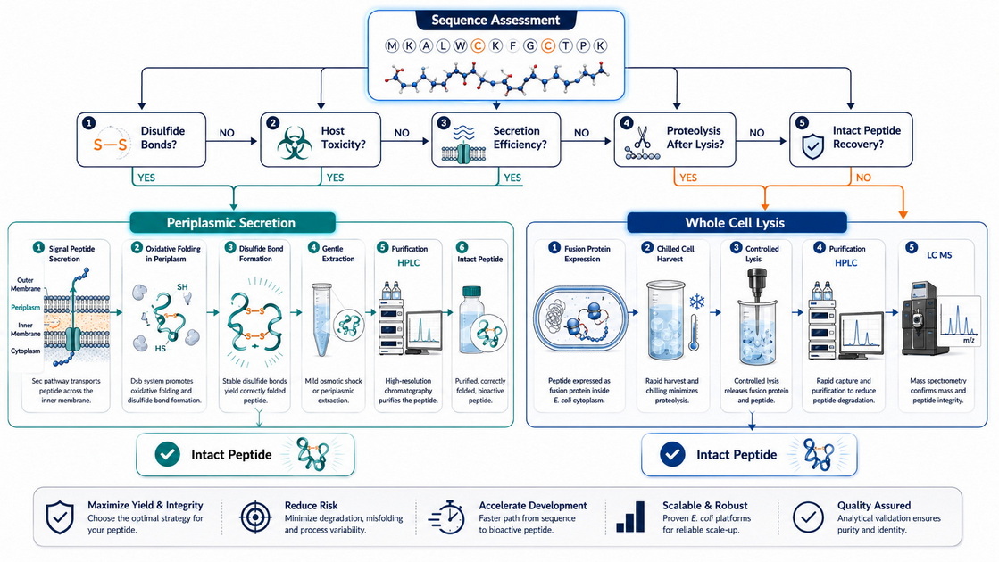
A Practical Decision Workflow for Peptide Expression
Rather than committing to one route based only on literature precedent, a focused parallel screen is usually more informative.
1: Assess the Peptide Sequence
Review the following properties before selecting a construct:
- Number and pattern of cysteines.
- Predicted disulfide-bond dependency.
- Net charge and hydrophobicity.
- Presence of protease-sensitive motifs.
- Likelihood of antimicrobial, cytotoxic, or membrane-active behavior.
- Need for an authentic N-terminus.
- Sensitivity to terminal additions after cleavage.
A peptide with multiple nonconsecutive cysteines and a defined disulfide framework is a strong candidate for early periplasmic screening. A highly toxic antimicrobial peptide may be better evaluated first as a protected intracellular fusion construct.
2: Build a Small, Purposeful Construct Panel
For many projects, a three-to-six construct panel can generate more useful information than repeated optimization of a single design.
A practical panel may include:
- Cytoplasmic fusion expression.
- Periplasmic fusion expression with one signal peptide.
- Periplasmic fusion expression with an alternative signal peptide.
- Different linker or cleavage-site configurations.
- Optional host-strain comparison where protease-related performance is suspected.
Avoid evaluating only total expression level. A strong band on a gel may not represent intact, active, correctly processed peptide.
3: Measure Intact Product, Not Just Apparent Yield
For small peptides, analytical confirmation should distinguish intact product from fragments, precursors, dimers, mispaired disulfide forms, and fusion-derived impurities.
A practical analytical package may include:
- Reducing and non-reducing electrophoretic analysis where applicable.
- Reversed-phase HPLC for purity and degradation profiles.
- LC-MS or intact-mass analysis to confirm molecular identity.
- Peptide mapping when sequence clipping or disulfide status is a concern.
- Functional assays matched to the peptide's intended research use.
This is particularly important because a peptide can appear "expressed" while actually existing as a mixture of truncated or improperly processed species.
4: Use Time-Course Sampling
Time-course data can identify whether degradation occurs during expression, harvest, extraction, or purification.
For example, collect samples at:
1. Pre-induction.
2. Early induction.
3. Mid-induction.
4. End of induction.
5. Immediately after harvest.
6. Immediately after extraction or lysis.
7. After clarification.
8. After the first capture step.
If intact peptide declines sharply after lysis but not during expression, the bottleneck is likely downstream exposure rather than intracellular synthesis. If degradation occurs before harvest, construct design, host compatibility, induction strength, or compartment targeting should be reconsidered.
5: Select the Route Based on Total Usable Recovery
The most valuable metric is not total cellular peptide. It is intact, correctly characterized peptide recovered after purification.
A lower-expression periplasmic construct may outperform a high-expression cytoplasmic construct if it produces a cleaner, more stable, more readily purified product. Conversely, a high-performing fusion construct recovered by rapid whole-cell lysis may be the better choice if secretion creates major transport losses or processing heterogeneity.
Reducing Proteolysis Before It Becomes a Downstream Problem
In practice, proteolysis is easiest to control before the peptide is released as a free product. This leads to several design principles.
Design for Protection, Then Release
For vulnerable peptides, consider protecting the sequence during expression and liberating it later through a controlled cleavage strategy. This may involve a fusion partner, a protease-recognition site, or a self-processing design. The critical point is to confirm that cleavage produces the required peptide termini and does not introduce unwanted residues.
Avoid Overdriving Expression
Very strong expression can overwhelm folding, secretion, and quality-control systems. The result may be aggregation, stress responses, incomplete export, or proteolysis. Moderate expression can sometimes yield more intact peptide than maximal induction.
Treat Signal Peptides as Screening Variables
Signal peptides are not interchangeable. Their translocation kinetics, cleavage behavior, and compatibility with the mature sequence can differ. A signal peptide that performs well for one recombinant protein may not perform well for another. Published comparisons have shown that DsbA-derived signal sequences can enhance soluble and functional production for some targets, but this must be verified experimentally for each peptide.
Confirm Sequence Integrity Early
For small bioactive peptides, mass-based confirmation should be introduced early in development. It is far more efficient to detect clipping after a small-scale screen than after scale-up and extended purification work.
Conclusion
For small bioactive peptides in E. coli, periplasmic secretion and whole-cell lysis solve different parts of the same stability problem.
Periplasmic secretion can be especially valuable when a peptide needs an oxidative environment for disulfide formation, when cytoplasmic degradation is severe, or when a cleaner initial extract can improve recovery. Successful export may reduce exposure to some cytoplasmic proteases, but it still requires careful attention to signal-peptide selection, translocation efficiency, and periplasmic degradation.
Whole-cell lysis remains a powerful and flexible route, especially for peptides expressed as protected fusion proteins or sequences that cannot be exported efficiently. Its major risk is the rapid mixing of the peptide with a complex pool of host proteases after disruption. Fast, cold, selective, and analytically controlled recovery is therefore essential.
For peptide expression programs, the most reliable strategy is often a parallel, evidence-driven comparison: screen periplasmic and cytoplasmic-fusion formats, monitor intact molecular species over time, and select the route that delivers the highest recovery of correctly characterized material for early discovery and characterization.
Gene Universal can support research teams with end-to-end DNA/RNA-to-protein and antibody expression workflows, including construct design, recombinant expression strategy evaluation, peptide and protein characterization planning, and fit-for-purpose research-grade material generation. Contact our scientific team to discuss an E. coli peptide-expression strategy tailored to your sequence, structural requirements, and research goals.
Frequently Asked Questions
1. Is periplasmic secretion always better than whole-cell lysis for small peptides?
No. Periplasmic secretion can reduce exposure to some cytoplasmic proteases and support disulfide formation, but export can be inefficient or sequence dependent. Whole-cell lysis may perform better for peptides that require fusion-based protection, are poorly translocated, or are difficult to recover from the periplasm.
2. Why are small bioactive peptides more susceptible to proteolytic degradation?
Small peptides often have exposed termini and flexible, accessible cleavage sites. They may lack the stable folded core that protects larger proteins. Some bioactive peptides can also stress or damage the host cell, which further complicates expression.
3. Can a fusion partner reduce peptide degradation in E. coli?
Yes. Fusion expression is commonly used to improve recombinant peptide stability, reduce host toxicity, and protect the peptide from intracellular proteolysis. The fusion partner, linker, and cleavage method should be selected carefully to preserve the correct final peptide sequence.
4. Does the E. coli periplasm help form disulfide bonds?
Often, yes. The periplasm provides an oxidizing environment and contains protein-folding components associated with disulfide-bond formation. This makes it a useful compartment for many cysteine-rich peptide and protein targets.
5. How can I determine whether degradation happens during expression or after lysis?
Use a timed sampling plan that includes samples during induction, at harvest, immediately after lysis or periplasmic extraction, after clarification, and after initial purification. Analyze intact mass and chromatographic profiles to locate when peptide loss begins.
6. Which analytical methods are most useful for checking peptide integrity?
Reversed-phase HPLC and LC-MS are especially valuable because they can reveal truncation, clipping, oxidation, and mass heterogeneity. For disulfide-containing peptides, non-reducing analysis, peptide mapping, and functional assays can provide additional confirmation.
7. Can lower expression temperature reduce peptide degradation?
It can help in some systems by reducing expression stress, improving folding, or changing protease-related behavior. However, the outcome depends on the peptide, host strain, construct, fusion partner, and induction conditions. Temperature should be treated as a screening variable rather than a universal solution.
References
1. Gottesman S, Clark WP, de Crecy-Lagard V, Maurizi MR. (https://pubmed.ncbi.nlm.nih.gov/2644253/).
2. Baneyx F, Georgiou G. [Purification of secreted recombinant proteins from (https://pubmed.ncbi.nlm.nih.gov/1367083/).
3. Li Y. (https://pubmed.ncbi.nlm.nih.gov/21843642/).
4. Parachin NS, Mulder KC, Viana AA, Dias SC, Franco OL. (https://pubmed.ncbi.nlm.nih.gov/20094858/).
5. de Marco A. (https://pubmed.ncbi.nlm.nih.gov/28470603/).
6. Hatahet F, Boyd D, Beckwith J. (https://pubmed.ncbi.nlm.nih.gov/21241169/).
7. Mavridou DAI, Ferguson SJ, Stevens JM. (https://pubmed.ncbi.nlm.nih.gov/31396164/).
8. Baneyx F. (https://pubmed.ncbi.nlm.nih.gov/8812844/).
9. Sugimoto S, Yamanaka K, Nishikori S, et al. (https://pubmed.ncbi.nlm.nih.gov/3277950/).
10. Li Y, Wang H, Lv Z, et al. (https://pubmed.ncbi.nlm.nih.gov/33656890/).